publications
An up-to-date list is available on Google Scholar.
2025
-
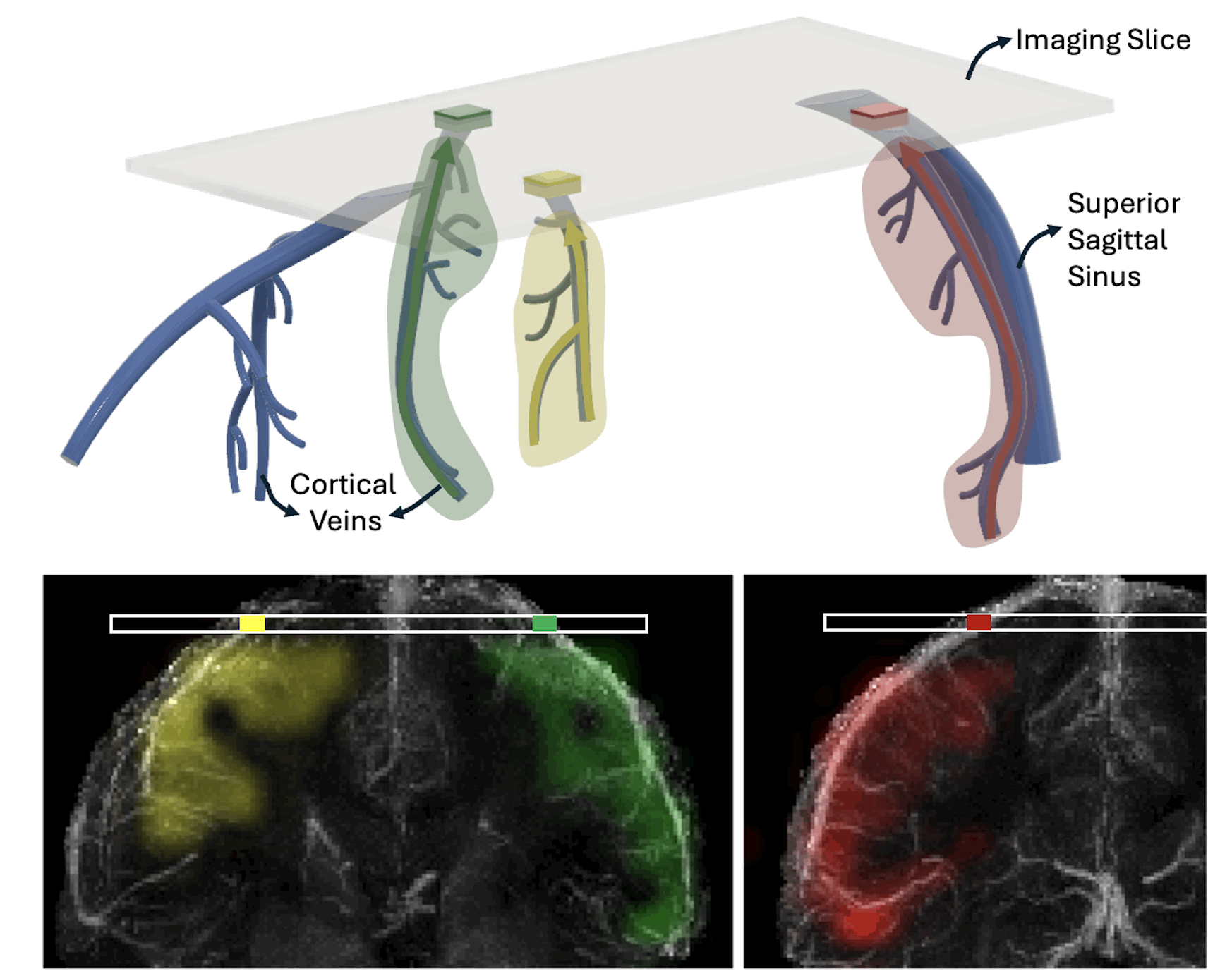 MR perfusion source mapping depicts venous territories and reveals perfusion modulation during neural activationEkin Karasan, Jingjia Chen, Julian Maravilla, and 3 more authorsNature Communications, 2025
The cerebral venous system plays a crucial role in neurological and vascular conditions, yet its hemodynamics remain underexplored due to its complexity and variability across individuals. To address this, we develop a venous perfusion source mapping method using Displacement Spectrum MRI, a non-contrast technique that leverages blood water as an endogenous tracer. Our technique encodes spatial information into the magnetization of blood water spins during tagging and detects it once the tagged blood reaches the brain’s surface, where the signal-to-noise ratio is 3–4 times higher. We resolve the sources of blood entering the imaging slice across short (10 ms) to long (3 s) evolution times, effectively capturing perfusion sources in reverse. This approach enables the measurement of slow venous blood flow, including potential contributions from capillary beds and surrounding tissue. We demonstrate perfusion source mapping in the superior cerebral veins, verify its sensitivity to global perfusion modulation induced by caffeine, and establish its specificity by showing repeatable local perfusion modulation during neural activation. From all blood within the imaging slice, our method localizes the portion originating from an activated region upstream.
2024
- ThesisVenous Perfusion Source Mapping “in Reverse” with Magnetic Resonance ImagingEkin KarasanUniversity of California, Berkeley, 2024
2023
-
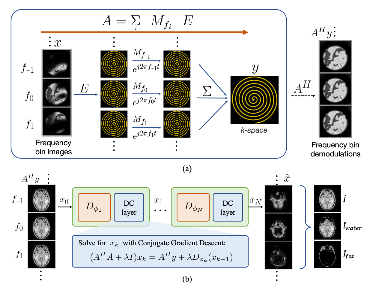 ResoNet: a Physics-Informed DL Framework for Off-Resonance Correction in MRI Trained with NoiseAlfredo De Goyeneche, Shreya Ramachandran, Ke Wang, and 4 more authorsIn Thirty-seventh Conference on Neural Information Processing Systems, 2023
Magnetic Resonance Imaging (MRI) is a powerful medical imaging modality that offers diagnostic information without harmful ionizing radiation. Unlike optical imaging, MRI sequentially samples the spatial Fourier domain k-space of the image. Measurements are collected in multiple shots, or readouts, and in each shot, data along a smooth trajectory is sampled. Conventional MRI data acquisition relies on sampling k-space row-by-row in short intervals, which is slow and inefficient. More efficient, non-Cartesian sampling trajectories (e.g., Spirals) use longer data readout intervals, but are more susceptible to magnetic field inhomogeneities, leading to off-resonance artifacts. Spiral trajectories cause off-resonance blurring in the image, and the mathematics of this blurring resembles that of optical blurring, where magnetic field variation corresponds to depth and readout duration to aperture size. Off-resonance blurring is a system issue with a physics-based, accurate forward model. We present a physics-informed deep learning framework for off-resonance correction in MRI, which is trained exclusively on synthetic, noise-like data with representative marginal statistics. Our approach allows for fat/water partial volume effects modeling and separation, and parallel imaging acceleration. Through end-to-end training using synthetic randomized data (i.e., images, coil sensitivities, field maps), we train the network to reverse off-resonance effects across diverse anatomies and contrasts without retraining. We demonstrate the effectiveness of our approach through results on phantom and in-vivo data. This work has the potential to facilitate the clinical adoption of non-Cartesian sampling trajectories, enabling efficient, rapid, and motion-robust MRI scans. Code is publicly available at: https://github.com/mikgroup/ResoNet
-
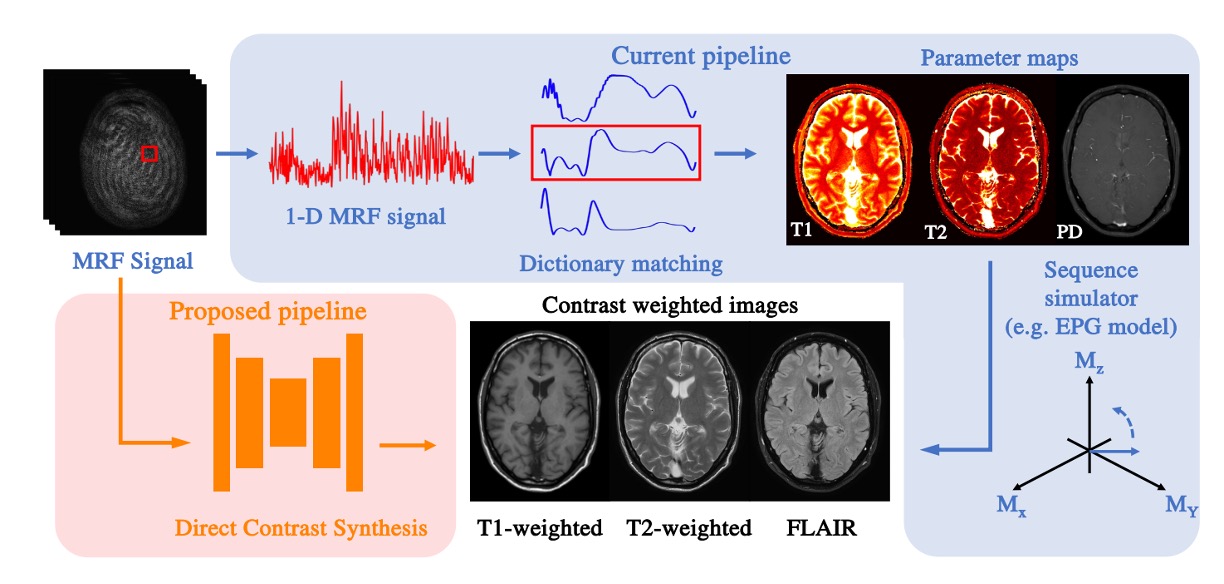 High-fidelity direct contrast synthesis from magnetic resonance fingerprintingKe Wang, Mariya Doneva, Jakob Meineke, and 6 more authorsMagnetic Resonance in Medicine, 2023
Magnetic Resonance Fingerprinting (MRF) is an efficient quantitative MRI technique that can extract important tissue and system parameters such as T1, T2, B0, and B1 from a single scan. This property also makes it attractive for retrospectively synthesizing contrast-weighted images. In general, contrast-weighted images like T1-weighted, T2-weighted, etc., can be synthesized directly from parameter maps through spin-dynamics simulation (i.e., Bloch or Extended Phase Graph models). However, these approaches often exhibit artifacts due to imperfections in the mapping, the sequence modeling, and the data acquisition. Here we propose a supervised learning-based method that directly synthesizes contrast-weighted images from the MRF data without going through the quantitative mapping and spin-dynamics simulation. To implement our direct contrast synthesis (DCS) method, we deploy a conditional Generative Adversarial Network (GAN) framework and propose a multi-branch U-Net as the generator. The input MRF data are used to directly synthesize T1-weighted, T2-weighted, and fluid-attenuated inversion recovery (FLAIR) images through supervised training on paired MRF and target spin echo-based contrast-weighted scans. In-vivo experiments demonstrate excellent image quality compared to simulation-based contrast synthesis and previous DCS methods, both visually as well as by quantitative metrics. We also demonstrate cases where our trained model is able to mitigate in-flow and spiral off-resonance artifacts that are typically seen in MRF reconstructions and thus more faithfully represent conventional spin echo-based contrast-weighted images.
-
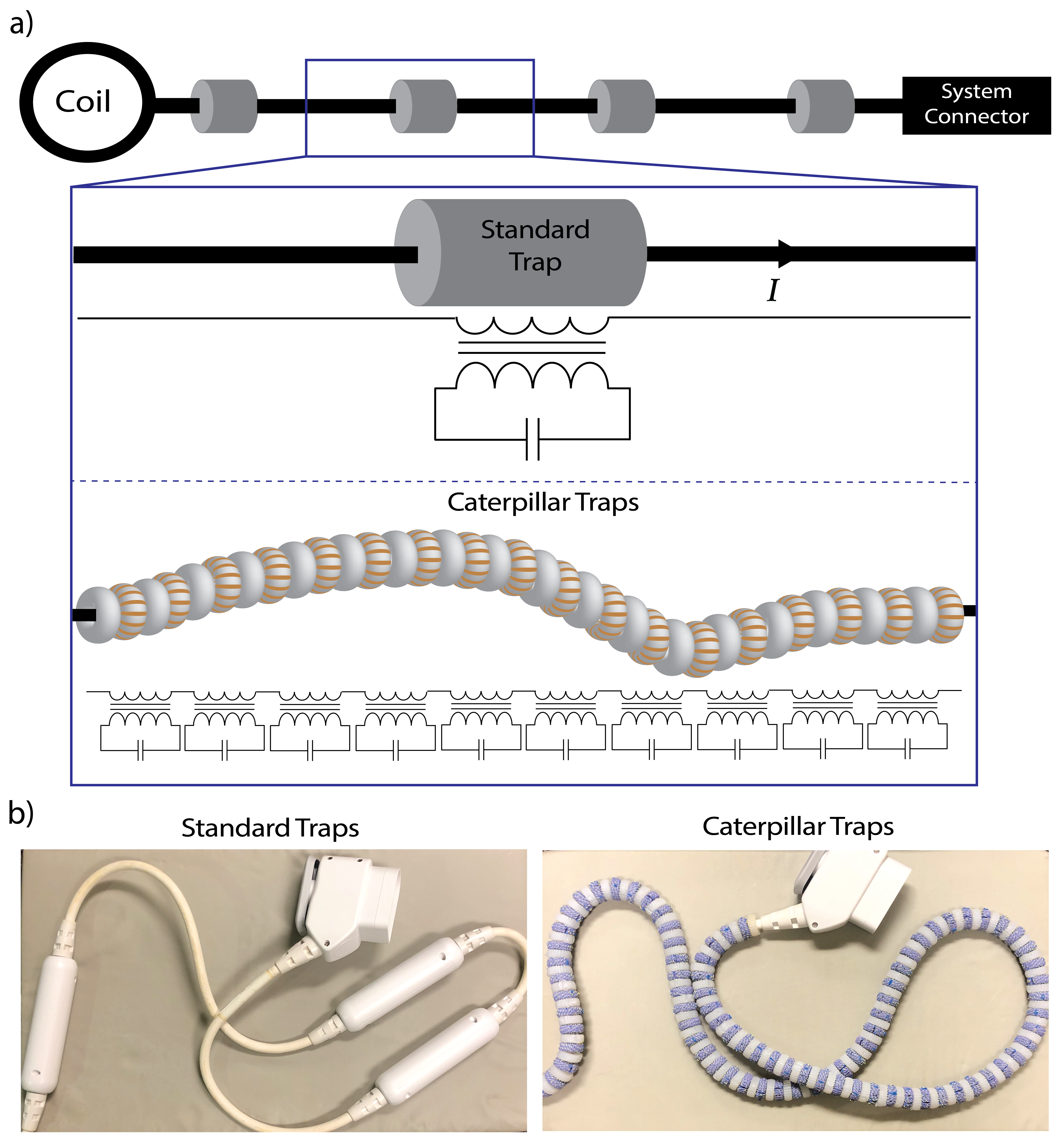 Caterpillar traps: A highly flexible, distributed system of toroidal cable trapsEkin Karasan, Alison Hammerschmidt, Ana C Arias, and 3 more authorsMagnetic resonance in medicine, 2023
Purpose: Coil arrays are connected to the main MRI system with long, shielded coaxial cables. RF coupling of these cables to the main transmit coil can cause high shield currents, which pose risks of heating and RF burns. High-blocking resonant RF traps are placed at distinct positions along cables to mitigate these currents. Traditional traps are designed to be stiff to avoid changes in their resonant frequency, hindering the overall system flexibility. Instead of using a few high-blocking traps, we propose the use of caterpillar traps—a distributed system of small, elastic traps that cover the full length of cables. Methods: We leverage an array of resonant toroids as traps, forming a caterpillar-like structure whereby bending only impacts individual traps minimally. Benchtop measurements are used to determine the blocking of caterpillar traps and show their robustness to bending. We also compare an anterior array system cable covered with caterpillar traps to a commercial cable with B1+ and heating measurements. Results: Benchtop experiments with caterpillar traps demonstrate high robustness to bending. B1+ mapping experiments of an anterior array cable show improved blocking and flexibility compared to a commercial cable. Conclusion: Caterpillar traps provide sufficient attenuation to shield currents while allowing cable flexibility. Our distributed design can provide high blocking efficiency at different positions and orientations, even in cases where commercial cable traps cannot.
-
 High-resolution multi-shot diffusion-weighted MRI combining markerless prospective motion correction and locally low-rank constrained reconstructionHao Chen, Ke Dai, Sijie Zhong, and 8 more authorsMagnetic resonance in medicine, 2023
Purpose: Subject head motion is a major challenge in DWI, leading to image blurring, signal losses, and biases in the estimated diffusion parameters. Here, we investigate a combined application of prospective motion correction and spatial-angular locally low-rank constrained reconstruction to obtain robust, multi-shot, high-resolution diffusion-weighted MRI under substantial motion. Methods: Single-shot EPI with retrospective motion correction can mitigate motion artifacts and resolve any mismatching of gradient encoding orientations; however, it is limited by low spatial resolution and image distortions. Multi-shot acquisition strategies could achieve higher resolution and image fidelity but increase the vulnerability to motion artifacts and phase variations related to cardiac pulsations from shot to shot. We use prospective motion correction with optical markerless motion tracking to remove artifacts and reduce image blurring due to bulk motion, combined with locally low-rank regularization to correct for remaining artifacts due to shot-to-shot phase variations. Results: The approach was evaluated on healthy adult volunteers at 3 Tesla under different motion patterns. In multi-shot DWI, image blurring due to motion with 20 mm translations and 30° rotations was successfully removed by prospective motion correction, and aliasing artifacts caused by shot-to-shot phase variations were addressed by locally low-rank regularization. The ability of prospective motion correction to preserve the orientational information in DTI without requiring a reorientation of the b-matrix is highlighted. Conclusion: The described technique is proved to hold valuable potential for mapping brain diffusivity and connectivity at high resolution for studies in subjects/cohorts where motion is common, including neonates, pediatrics, and patients with neurological disorders.
2021
-
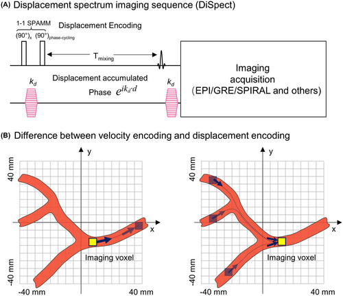 DiSpect: Displacement spectrum imaging of flow and tissue perfusion using spin-labeling and stimulated echoesZhiyong Zhang, Ekin Karasan, Karthik Gopalan, and 2 more authorsMagnetic Resonance in Medicine, 2021
Purpose: We propose a new method, displacement spectrum (DiSpect) imaging, for probing in vivo complex tissue dynamics such as motion, flow, diffusion, and perfusion. Based on stimulated echoes and image phase, our flexible approach enables observations of the spin dynamics over short (milliseconds) to long (seconds) evolution times. Methods: The DiSpect method is a Fourier‐encoded variant of displacement encoding with stimulated echoes, which encodes bulk displacement of spins that occurs between tagging and imaging in the image phase. However, this method fails to capture partial volume effects as well as blood flow. The DiSpect variant mitigates this by performing multiple scans with increasing displacement‐encoding steps. Fourier analysis can then resolve the multidimensional spectrum of displacements that spins exhibit over the mixing time. In addition, repeated imaging following tagging can capture dynamic displacement spectra with increasing mixing times. Results: We demonstrate properties of DiSpect MRI using flow phantom experiments as well as in vivo brain scans. Specifically, the ability of DiSpect to perform retrospective vessel‐selective perfusion imaging at multiple mixing times is highlighted. Conclusion: The DiSpect variant is a new tool in the arsenal of MRI techniques for probing complex tissue dynamics. The flexibility and the rich information it provides open the possibility of alternative ways to quantitatively measure numerous complex spin dynamics, such as flow and perfusion within a single exam.
2020
- IEEEComputational MRI with physics-based constraints: Application to multicontrast and quantitative imagingJonathan I Tamir, Frank Ong, Suma Anand, and 3 more authorsIEEE signal processing magazine, 2020
Compressed sensing (CS) takes advantage of a low-dimensional signal structure to reduce sampling requirements to far below the Nyquist rate. In magnetic resonance imaging (MRI), this often takes the form of sparsity through wavelet transforms, finite differences, and low-rank extensions. Though powerful, these image priors are phenomenological in nature and do not account for the mechanism behind the image formation. On the other hand, MRI signal dynamics are governed by physical laws, which can be explicitly modeled and used as priors for reconstruction. These explicit and implicit signal priors can be synergistically combined in an inverse-problem framework to recover sharp, multicontrast images from highly accelerated scans. Furthermore, the physics-based constraints provide a recipe for recovering quantitative, biophysical parameters from the data.
2018
-
 An enhanced mechanistic model for capnography, with application to CHF-COPD discriminationEkin Karasan, Abubakar Abid, Rebecca J Mieloszyk, and 3 more authorsIn 2018 40th Annual International Conference of the IEEE Engineering in Medicine and Biology Society (EMBC), 2018
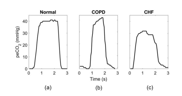
Capnography records CO2 partial pressure in exhaled breath as a function of time or exhaled volume. Time-based capnography, which is our focus, is a point-of-care, noninvasive, effort-independent and widely available clinical monitoring modality. The generated waveform, or capnogram, reflects the ventilation-perfusion dynamics of the lung, and thus has value in the diagnosis of respiratory conditions such as chronic obstructive pulmonary disease (COPD). Effective discrimination between normal respiration and obstructive lung disease can be performed using capnogram-derived estimates of respiratory parameters in a simple mechanistic model of CO2 exhalation. We propose an enhanced mechanistic model that can capture specific capnogram characteristics in congestive heart failure (CHF) by incorporating a representation of the inertance associated with fluid in the lungs. The 4 associated parameters are estimated on a breath-by-breath basis by fitting the model output to the exhalations in the measured capnogram. Estimated parameters from 40 exhalations of 7 CHF and 7 COPD patients were used as a training set to design a quadratic discriminator in the parameter space, aimed at distinguishing between CHF and COPD patients. The area under the ROC curve for the training set was 0.94, and the corresponding equal-error-rate value of approximately 0.1 suggests classification accuracies of the order of 90% are attainable. Applying this discriminator without modification to 40 exhalations from each CHF and COPD patient in a fresh test set, and deciding on a simple majority basis whether the patient has CHF or COPD, results in correctly labeling all 8 out of the 8 CHF patients and 6 out of the 8 COPD patients in the test set, corresponding to a classification accuracy of 87.5%.
2017
-
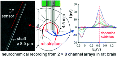 Subcellular probes for neurochemical recording from multiple brain sitesHelen N Schwerdt, Min Jung Kim, Satoko Amemori, and 8 more authorsLab on a Chip, 2017
Dysregulation of neurochemicals, in particular, dopamine, is epitomized in numerous debilitating disorders that impair normal movement and mood aspects of our everyday behavior. Neurochemical transmission is a neuron-specific process, and further exhibits region-specific signaling in the brain. Tools are needed to monitor the heterogeneous spatiotemporal dynamics of dopamine neurotransmission without compromising the physiological processes of the neuronal environment. We developed neurochemical probes that are ten times smaller than any existing dopamine sensor, based on the size of the entire implanted shaft and its sensing tip. The microfabricated probe occupies a spatial footprint (9 μm) coordinate with the average size of individual neuronal cells (∼10 μm). These cellular-scale probes were shown to reduce inflammatory response of the implanted brain tissue environment. The probes are further configured in the form of a microarray to permit electrochemical sampling of dopamine and other neurotransmitters at unprecedented spatial densities and distributions. Dopamine recording was performed concurrently from up to 16 sites in the striatum of rats, revealing a remarkable spatiotemporal contrast in dopamine transmission as well as site-specific pharmacological modulation. Collectively, the reported platform endeavors to enable high density mapping of the chemical messengers fundamentally involved in neuronal communication through the use of minimally invasive probes that help preserve the neuronal viability of the implant environment.