Venous Perfusion Source Mapping
A non-contrast MRI technique to trace blood flow and map venous territories.
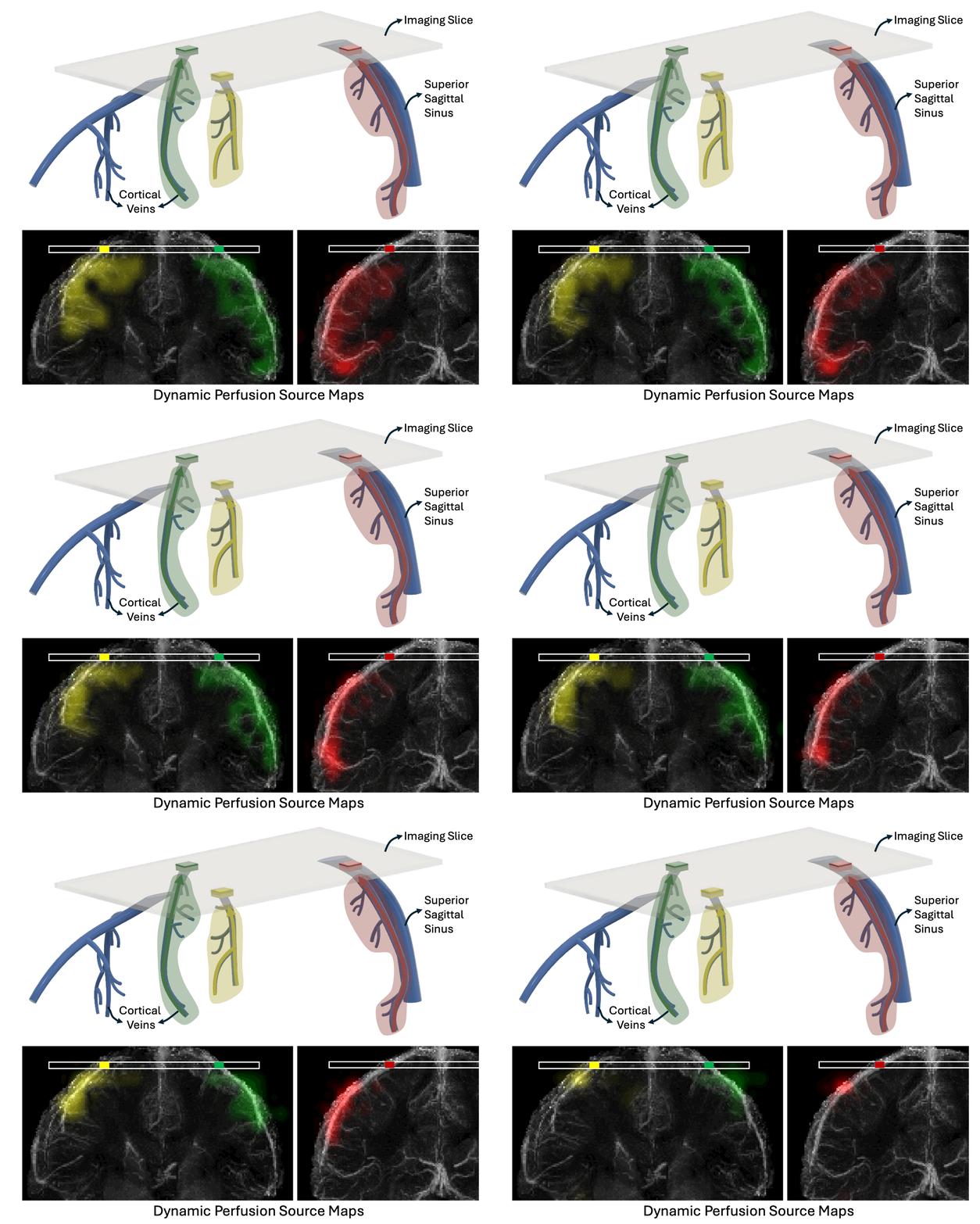
Despite its importance to neurological and vascular disease, venous cerebral hemodynamics remain underexplored because of their complexity and person-to-person variability. We developed a novel MRI method that traces venous blood “in reverse”, revealing the perfusion, or blood flow, sources of different cerebral veins. Using water in blood as a natural tracer, we initially tag the blood with information that lasts for a few seconds as the blood drains from capillaries into larger veins. When that blood reaches the bigger veins, we decode the tagged information to determine where the blood came from.

Our approach is based on Displacement Spectrum MRI (DiSpect): we encode spatial information into the magnetization of blood-water spins during tagging and read it out once the tagged blood enters the imaging slice at the brain surface, where the signal-to-noise ratio is ~3–4× higher. By sampling at short-to-long evolution times (10 ms to ~2 s), we resolve the sources of blood entering an imaging slice, capturing slow venous flow with potential contributions from capillaries and surrounding tissue.

Perfusion source mapping detects global modulation in perfusion (e.g., due to caffeine) and shows specificity to localized perfusion changes due to functional activation. Remarkably, within the blood arriving to a single image voxel, our method localizes the blood originating from an activated region upstream. For details, see our article in Nature Communications and for a broader overview, see the recent feature by Berkeley Engineering News.